
29 September 2026
When to choose minimally invasive pain treatments
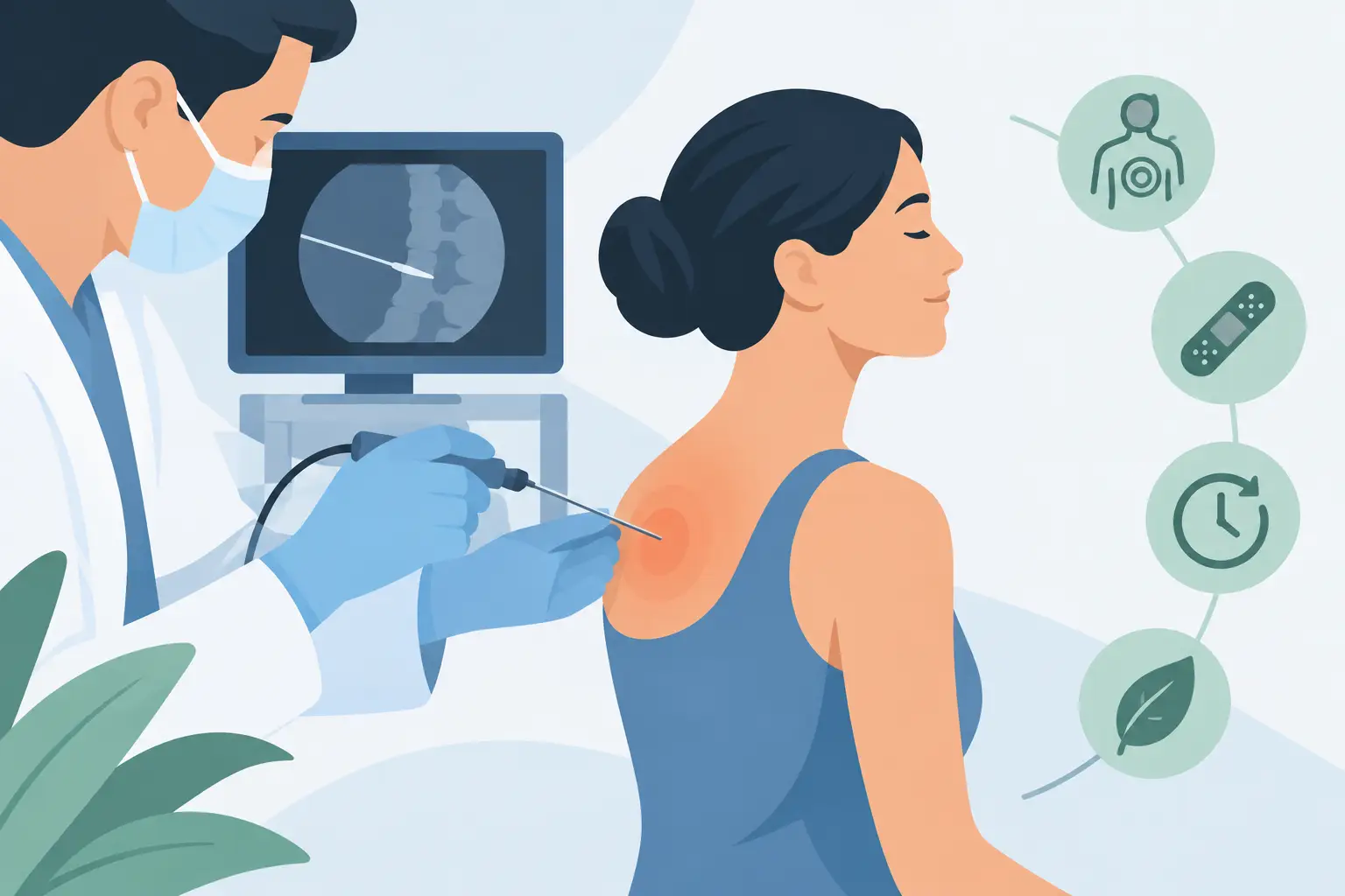
Shoulder pain that stops you getting dressed, a knee that makes stairs difficult or low back pain that returns without fail after every working day do not always require surgery. In selected cases, minimally invasive pain treatments make it possible to act on the spot responsible for the symptoms through the skin, using fine needles and ultrasound or radiological guidance. The aim is not simply to “give an injection”: it is to identify precisely the structure involved, treat it while preserving healthy tissue and build a realistic recovery.
What minimally invasive treatment means
Minimally invasive treatment means a procedure performed without a traditional surgical incision, or through a skin access of just a few millimetres. Imaging - especially ultrasound, X-ray or angiography - makes it possible to see needles, tendons, joints, vessels and calcium deposits in real time. This reduces guesswork and makes it possible to reach a deep target without needlessly crossing other structures.
In the field of musculoskeletal pain, procedures may include targeted ultrasound-guided injections, percutaneous lavage of shoulder calcifications, treatment of certain tendon and joint conditions, or diagnostic and therapeutic manoeuvres that help understand how much a specific structure contributes to the pain. In other areas, such as the pelvis, pain may be due to vascular or gynaecological abnormalities and require a different interventional assessment, for example embolization.
“Minimally invasive” does not mean automatic, risk-free or suitable for everyone. It means choosing an approach proportionate to the problem, when the clinical indications make it reasonable.
Minimally invasive pain treatments: when they may be indicated
The first useful question is not “which procedure should I have?”, but “where is the pain really coming from?”. An MRI or X-ray report may show changes that are common even in people without symptoms. Conversely, very limiting pain may be due to inflammation or a mechanical impingement, and imaging must be interpreted together with the clinical examination.
A targeted procedure may be considered when pain persists despite an adequate course of conservative treatment, when it prevents physiotherapy and movement, or when there is a clearly identifiable anatomical target. These are common situations in calcific tendinopathy of the shoulder, resistant epicondylitis, some forms of plantar fasciitis, painful osteoarthritis of the thumb or wrist, hip and knee pain and selected cases of chronic low back pain.
The duration of symptoms, however, is not enough. What matters is the type of pain, the loss of function, the treatments already tried, work or sporting activity, the medicines being taken and general health. Fever, significant trauma, sudden loss of strength, loss of sensation, bladder or bowel disturbances or marked swelling, on the other hand, require urgent assessment and must not be managed as simple inflammatory pain.
A practical example: shoulder calcification
In the shoulder, a tendon calcification may remain silent or cause intense pain, often at night, and limit raising the arm. If the symptoms are consistent with the location of the calcification and conservative treatment is not enough, ultrasound-guided percutaneous lavage, also called lithoclasty or barbotage, may be an option.
During the procedure the doctor visualises the deposit on ultrasound, introduces fine needles and breaks it up or washes it out with saline solution when its consistency and position allow. The aim is to reduce the calcific material and local irritation, not to surgically “repair” the tendon. Some calcifications - very small, hard or at a particular stage of their evolution - may not be suitable for lavage. In these cases rehabilitation, drug therapy, shockwave therapy or, more rarely, surgery may be preferable.
How an image-guided procedure is carried out
The pathway begins with a specialist consultation. Symptoms, existing tests, treatments received and the patient’s expectations are gathered. Ultrasound performed during the assessment, when indicated, does not just confirm a suspicion: it makes it possible to correlate the image with the exact spot of the pain and with the movement that triggers it.
Before treatment you need to report allergies, anticoagulant or antiplatelet therapy, diabetes, pregnancy, current infections and previous reactions to drugs or contrast media. Stopping medicines that affect clotting must never be decided on your own: it depends on the risk of the procedure and on the reason those medicines were prescribed.
Most musculoskeletal injections and lavages use local anaesthesia or a very limited form of anaesthesia. The patient stays awake, can report unusual sensations and, unless advised otherwise, goes home the same day. Vascular procedures such as some embolizations require more structured preparation and monitoring, but still avoid a traditional surgical incision and often general anaesthesia.
After treatment, a temporary increase in local pain may occur, particularly in the first 24–72 hours. This is a possibility to be aware of beforehand, not an automatic sign that the procedure has not worked. Relative rest, ice when appropriate and prescribed medication help manage this phase. Return to work depends on the site treated and the type of job: sedentary work can resume soon, whereas lifting, sport or repetitive movements require a more cautious timeline.
Precision does not mean a promise of immediate recovery
The advantage of image guidance is precision: the drug or instrument is placed where it is needed, avoiding treating a nearby area “blind”. This can reduce unnecessary exposure of tissues and increase the technical reliability of the procedure. It does not, however, remove the biological limits of the condition.
An injection can ease inflammation and make physiotherapy possible again, but on its own it does not correct muscle weakness, work-related overload or a postural abnormality. Lavage of a calcification can improve pain and mobility, but the tendon must then recover in its own time. In the osteoarthritic knee, targeted treatments can reduce symptoms in selected patients, without recreating lost cartilage or always replacing a joint replacement when osteoarthritis is advanced.
Results also vary. They depend on the diagnosis, the stage of the disease, the quality of the tissue, daily habits and adherence to the rehabilitation programme. Proper communication should indicate both the expected benefit and the possibility of a partial, temporary or absent response.
Risks, alternatives and shared decisions
Percutaneous procedures are generally less invasive than surgery, but they are not free of possible complications. Temporary pain, small haematomas, infection, reactions to drugs, bleeding and injury to nearby structures are events to be discussed according to the procedure and the site. Ultrasound or radiological guidance serves precisely to reduce these risks, but cannot eliminate them.
The alternative may be to continue with physiotherapy and load modification, use medication for defined periods, turn to shockwave therapy for specific indications or consider surgery. There is no hierarchy that applies to every patient. A shoulder calcification is not the same as a herniated disc, and pelvic pain from venous congestion is not approached in the same way as osteoarthritis of the thumb.
A well-made decision comes from concrete questions: what is the target to be treated? What benefit is realistic? How long does recovery take? What happens if you wait? Is there an equally effective conservative solution? The answer must be personalised, not a standard promise.
If pain persists and limits daily life, the useful step is to obtain an accurate diagnosis before choosing the quickest treatment. Sometimes the best solution will be an ultrasound-guided procedure; at other times it will be to avoid an unnecessary procedure and focus on the treatment best suited to the real cause of the problem.
Want to know if it applies to you?
At the first consultation we review your clinical history and previous imaging together.
More articles
The information on this page is for general education and does not replace a medical consultation. The indication for any treatment must always be assessed individually.