
5 October 2026
Embolization of arteriovenous malformations of the limbs
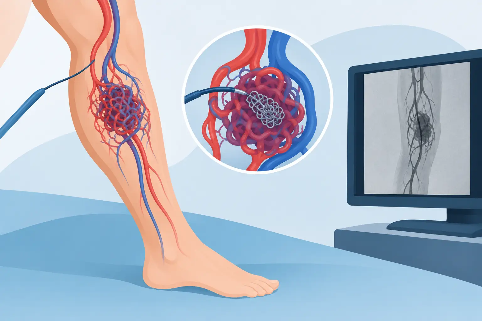
An arteriovenous malformation in a limb can cause pain, swelling, increased skin temperature, visible veins or a pulsating mass. In some cases it has been present since childhood but only becomes noticeable years later, perhaps following an injury, hormonal changes or progressive growth. Embolization of arteriovenous malformations of the limbs is a minimally invasive treatment that aims to selectively close the abnormal vessels responsible for the symptoms, while preserving healthy tissue as far as possible.
It is not, however, a standard procedure to be applied to every vascular lesion. Arteriovenous malformations (AVMs) are complex conditions: they require a precise diagnosis, image-based planning and a transparent comparison between embolization, surgery or a combined treatment.
What is an arteriovenous malformation of the limb
In normal circulation, blood passes from the arteries to the capillaries and then to the veins. In an AVM, instead, there is a network of abnormal connections, called the nidus, that links arteries and veins directly. High-pressure blood therefore reaches the veins without passing through the capillary bed.
This can cause the vessels to dilate, increase local blood flow and progressively infiltrate the tissues involved. An AVM can affect the hand, forearm, shoulder, foot, leg or thigh, and involve the skin, subcutaneous tissue, muscles, joints and, more rarely, bone. It should not be confused with a haemangioma, which is a vascular lesion with different biological characteristics and treatment.
Symptoms vary widely. Some malformations remain stable and do not require immediate treatment. Others cause pain at rest or on exertion, restricted movement, bleeding, skin ulcers, a feeling of heaviness, tingling or a difference in temperature compared with the opposite limb. When the lesion is very extensive and high-flow, it can also overload the general circulation.
When embolization of arteriovenous malformations of the limbs is indicated
The aim is not to treat a radiological image, but to address a real clinical problem: pain, bleeding, reduced function, growth of the lesion or risk of tissue damage. The decision depends on the location, the size, the vessels feeding the malformation and the presence of delicate structures nearby, such as nerves, tendons or joints.
Before treatment is proposed, colour Doppler ultrasound and contrast-enhanced MRI are usually needed. Colour Doppler ultrasound assesses the speed and direction of blood flow; MRI shows the extent of the lesion and its relationship with muscles, skin and deep structures. In many cases angiography, performed during the procedure, completes the map of the vessels to be treated.
Embolization can be the main option when surgery would involve a large incision, significant sacrifice of healthy tissue or a high functional risk. It can also precede surgery, reducing flow through the malformation and the risk of bleeding. In other situations, especially with diffuse lesions, the realistic aim is to control symptoms and slow progression, not necessarily to eliminate every single abnormal vessel in one session.
How the procedure is performed
Embolization is performed by an interventional radiologist in an angiography suite, under radiological guidance and, when useful, ultrasound guidance. After local anaesthesia and appropriate sedation if needed, a thin catheter is introduced through a small puncture, often in the groin, the wrist or directly into the malformation. No surgical incision is required.
Contrast medium is injected through the catheter to identify the feeding arteries, the nidus and the draining veins. This step is crucial: closing only an upstream artery, without precisely reaching the abnormal network, can encourage new feeding pathways to form over time.
Once the anatomy has been defined, embolic materials selected according to the type of flow and the location of the AVM are used. These may be liquid agents, particles, metal coils or dedicated devices. There is no single best material: the choice depends on how well the spread of the agent can be controlled, the size of the vessels and the need to protect arteries that supply healthy tissue.
The duration varies. Localised lesions can be treated in one session; complex or extensive AVMs may require procedures scheduled weeks or months apart. After treatment a compression dressing is applied to the access site and, in suitable cases, the patient goes home the same day or after a short period of observation.
What to expect in the following days
Local pain, swelling and a feeling of tightness in the treated area are possible and do not automatically indicate a complication. They often represent the inflammatory response to the closure of the abnormal vessels. Pain relief and advice on rest are tailored to the location and extent of the treatment.
Recovery does not always mean the lesion disappears immediately. The body needs to reabsorb or remodel the treated tissue, and improvement may be gradual. For an AVM of the hand or foot, for example, activities involving pressure or repeated strain may need to be resumed more cautiously than for a lesion elsewhere.
Real benefits and limitations to be aware of
The central advantage of embolization is precision: the treatment is guided by imaging and focused on the vessels responsible for the problem. This can avoid surgery or reduce its extent, limit scarring and, in many cases, allow treatment without general anaesthesia. The procedure also makes it possible to reach deep areas without extensively crossing muscles and skin.
The main limitation relates to the very nature of AVMs. These malformations have a marked ability to recruit new vessels over time, especially if the nidus is not treated sufficiently or if the lesion is highly infiltrative. For this reason, talking about a definitive cure without analysing the individual case would not be accurate. The result can be excellent in terms of pain, bleeding or function, but follow-up over time and, sometimes, further sessions may be needed.
Surgery also has a role. A well-defined AVM, after adequate devascularisation, can be removed with good results. Conversely, incomplete resection of a diffuse lesion can be complicated by recurrence and by a significant cosmetic or functional impact. The most appropriate strategy therefore comes from combining imaging, symptoms and the patient's goals.
Risks of embolization and warning signs to report
Like any vascular procedure, embolization carries risks that must be discussed before consent. Possible risks include a haematoma or bleeding at the puncture site, reaction to the contrast medium, infection, thrombosis and post-procedural pain. The most specific risk is unintended embolization of a healthy vessel, which can cause damage to the skin, muscles or nerves.
Careful planning, the use of thin catheters and continuous imaging control reduce these risks, but do not eliminate them. After returning home, rapidly increasing pain, fever, loss of sensation, cold or pale fingers or toes, persistent bleeding and the appearance of ulcers must be reported to the doctor promptly.
The right pathway: diagnosis, goal, follow-up
For an arteriovenous malformation of the limb, the useful question is not just “can it be embolized?”, but “what result is it reasonable to achieve in my case, and through what pathway?”. A well-indicated treatment starts by defining the priority symptom - pain, bleeding, difficulty walking or using the hand - and by a realistic assessment of the vascular anatomy.
After the procedure, clinical check-ups and colour Doppler ultrasound or MRI make it possible to confirm the reduction in flow and to plan any further treatment. Approaching an AVM in a targeted way means avoiding both passively waiting while symptoms progress and unrealistic promises: precision does not mean always doing more, but treating what is needed, at the right time, while preserving function and quality of life.
Want to know if it applies to you?
At the first consultation we review your clinical history and previous imaging together.
More articles
The information on this page is for general education and does not replace a medical consultation. The indication for any treatment must always be assessed individually.