
4 October 2026
Ultrasound-guided treatment of hip pain
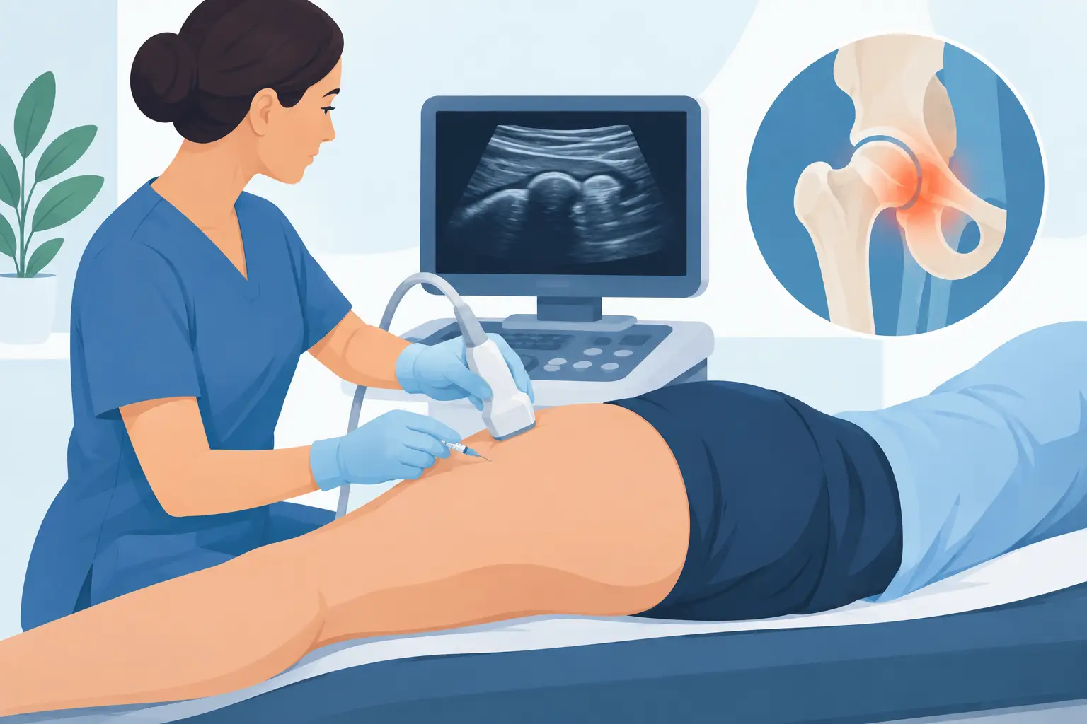
Tying your shoelaces, getting into the car, sleeping on your side or climbing the stairs: when your hip hurts, everyday movements can become limiting. Ultrasound-guided treatment of hip pain can be a targeted option when a specialist consultation has identified a specific structure responsible for the symptoms and conservative care has not been enough.
It is not a single procedure suitable for every type of hip pain. It is a way of performing injections and percutaneous treatments under ultrasound control, watching muscles, tendons, bursae and, in many cases, the path of the needle towards the target in real time. The aim is to treat the affected tissue more precisely, avoiding surgical incisions and, in appropriate outpatient procedures, without general anaesthesia and with discharge home the same day.
Where hip pain comes from
The term “hip” is often used for different areas. Pain in the groin may come from the hip joint itself, for example in the presence of osteoarthritis, synovitis or femoroacetabular impingement. Pain on the outer side of the hip is often associated with greater trochanteric pain syndrome, which can involve the trochanteric bursae and the gluteal tendons.
Pain at the back, near the buttock, may instead come from the lumbar spine, the sacroiliac joint, deep muscular structures or nerve irritation. Likewise, pain running down the thigh does not automatically mean a problem with the hip joint.
This distinction changes the treatment. Injecting a bursa when the real cause is lumbar radiculopathy does not solve the problem. That is why treatment should not start with the needle, but with a clinical assessment: the exact location of the pain, the movements that trigger it, how long symptoms have lasted, a physical examination and, if needed, X-rays, ultrasound or MRI.
When ultrasound-guided treatment of hip pain is indicated
Ultrasound provides a good view of many superficial and periarticular structures. It can therefore guide treatment of gluteal tendinopathies, trochanteric bursitis, enthesopathies, some iliopsoas conditions and other localised inflammatory conditions. It can also be useful for joint injections, depending on the clinical goal and the patient's anatomy.
Treatment is generally indicated when pain and functional limitation persist despite relative rest, activity modification, appropriate physiotherapy or medication prescribed by the doctor. In other cases, the injection also has diagnostic value: a significant, temporary reduction in pain after treating a suspected structure can help confirm where the symptoms are coming from.
Not every ultrasound finding requires a procedure. A minor tendon abnormality, for example, must be interpreted together with the symptoms reported by the patient and the consultation. Likewise, injection therapy does not replace regaining strength and pelvic control when these are part of the cause of overload.
How the procedure is performed
After checking the indications, available tests, current medications and possible contraindications, the doctor locates the structure to be treated with the ultrasound scanner. The skin is disinfected and the ultrasound image allows the needle to be followed as it advances, choosing the safest and most appropriate route.
The medication or substance used depends on the diagnosis. In some inflammatory conditions a local anaesthetic combined with an injectable anti-inflammatory drug may be indicated; in other cases the strategy may be different. There is no single “best” substance: the choice must take into account the location, the cause of the pain, the patient's clinical condition, previous treatments and functional goals.
The procedure usually takes a few minutes, although the appointment is longer because it includes assessment, planning and aftercare advice. Discomfort from the needle is usually limited; local anaesthesia can be used when indicated. For many injections neither hospital admission nor general anaesthesia is needed.
At the end a small dressing is applied. The patient receives personalised instructions on permitted activities, on managing pain in the following hours and on the signs that should prompt contacting the doctor. Same-day discharge is common, but this does not mean immediately returning to sport, long walks or physically demanding work.
What to expect after the injection
The response is not the same for everyone. If a local anaesthetic was used, there may be short-lived initial relief. The effect of the treatment on inflammation or pain may take a few days. Sometimes a temporary local flare-up is seen in the first 24-48 hours.
Recovery depends mainly on the condition treated. With bursitis or peritendinous irritation, it may be necessary to cut down for a few days on activities that compress or overload the area, such as sleeping on the painful side, running on slopes or repeatedly climbing stairs. With tendinopathies, the most lasting result often requires a progressive rehabilitation programme: load management, strengthening of the gluteal muscles and correction of the factors that contributed to the pain.
A well-performed injection can create a window of reduced pain that helps you move better again and make physiotherapy more effective. It does not automatically repair a degenerated tendon and does not eliminate osteoarthritis. Presenting it as a definitive solution would not be accurate.
Advantages of ultrasound guidance and real limitations
The main advantage of ultrasound is dynamic precision. The doctor can check the position of tendons, bursae, vessels and soft tissues, adapting the needle path to the individual anatomy. This is particularly relevant in the outer hip region, where the target can be very specific.
Ultrasound guidance reduces uncertainty compared with an injection performed using only external anatomical landmarks, but it does not make the treatment automatically necessary or risk-free. In addition, ultrasound has limitations in assessing some deep or intra-articular structures and, in selected circumstances, a different imaging technique or treatment pathway may be more appropriate.
How long the benefit lasts also varies. A patient with acute inflammation and modifiable loading may improve significantly; someone with advanced osteoarthritis, marked tendon degeneration or several sources of pain may obtain only partial or temporary benefit. If there is no effect, the diagnosis should be reassessed rather than repeating procedures without a clear clinical rationale.
Risks and precautions
Ultrasound-guided injections are minimally invasive procedures, but they are not free of possible side effects. Temporary pain at the entry point, bruising, bleeding and infection are possible, although uncommon with correct technique. Depending on the medication used, local or systemic effects may occur, and these should be discussed before the procedure.
Particular care is needed in the case of anticoagulant or antiplatelet therapy, diabetes, known allergies, fever or current infections, pregnancy and conditions that alter the risk of the procedure. Medication should not be stopped on your own initiative: its management must be decided by the doctor who knows your clinical situation and, when necessary, agreed with the prescribing specialist.
Hip pain that appears after a significant injury, or is associated with fever, sudden loss of strength, inability to bear weight or rapid worsening, requires prompt medical assessment, not simply an injection.
The most useful question is not whether an injection is “strong” or “weak”, but whether it is aimed at the structure that is truly causing the pain and part of a realistic treatment plan. When diagnosis, ultrasound guidance and rehabilitation work in the same direction, treatment can help restore movement with a targeted approach that respects the anatomy.
Want to know if it applies to you?
At the first consultation we review your clinical history and previous imaging together.
More articles
The information on this page is for general education and does not replace a medical consultation. The indication for any treatment must always be assessed individually.