
26 September 2026
Fibroid embolization: an alternative to hysterectomy
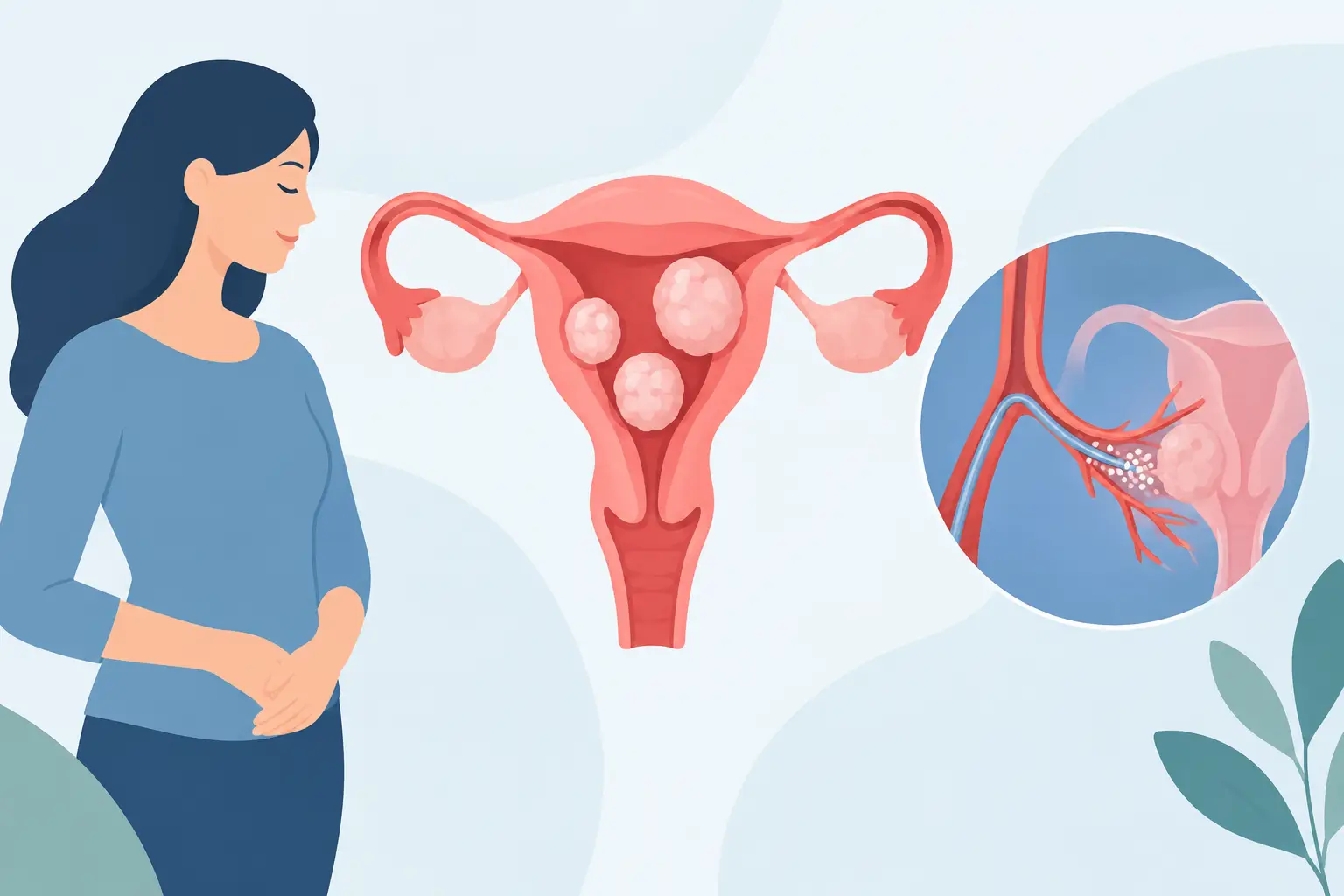
Menstrual bleeding so heavy that it affects your work, anaemia that keeps coming back despite iron, pressure in the lower abdomen or needing to pass urine frequently: these symptoms can make uterine fibroids much more than a simple finding on an ultrasound scan. Uterine fibroid embolization as an alternative to hysterectomy allows, in selected patients, the fibroids responsible for the symptoms to be treated while preserving the uterus and without a surgical incision.
It is neither an automatic choice nor a solution suitable for every fibroid. The decision requires a careful assessment of the symptoms, the location and size of the fibroids, age, plans for pregnancy and any treatments already tried. Understanding how it works helps you compare it with hysterectomy and the other options without oversimplifying.
Why fibroids may need treatment
Fibroids, or uterine myomas, are benign growths made of the muscle of the uterus. They are common and do not always cause symptoms. When they are small and symptom-free, regular gynaecological check-ups are often enough.
Things change when their position distorts the uterine cavity, increases the surface that bleeds during periods or presses on nearby organs. Submucosal fibroids, close to the inner cavity of the uterus, are often associated with heavy periods. Intramural or subserosal fibroids can cause pelvic heaviness, an enlarged abdomen, pain, constipation or urinary symptoms, especially when they are large.
Severity does not depend only on the centimetres given in the report. What counts is the real impact: how many days of work are lost, whether haemoglobin is low, whether pain limits daily activities and whether medication actually controls the symptoms. Treatment should be chosen for the person, not for the ultrasound image alone.
Uterine fibroid embolization: an alternative to hysterectomy
Embolization is an interventional radiology procedure that selectively reduces or blocks the blood supply to fibroids. The fibroids receive less oxygen and fewer nutrients, gradually shrink and, in most cases, the symptoms decrease.
The doctor inserts a very thin catheter through a small puncture, usually in the groin or wrist. Under radiological guidance, the catheter is advanced to the uterine arteries and microspheres designed to block the branches feeding the fibroids are released. This is not open surgery: no surgical incisions are made in the abdomen and, in most cases, general anaesthesia is not needed.
Image guidance is central to the procedure. It shows the path of the vessels, allows both sides of the uterus to be treated in a targeted way and makes it possible to check the technical result at the end of the embolization. The aim is not to physically remove every fibroid, but to reduce the blood supply that sustains their growth and the symptoms.
When it may be indicated
Embolization may be considered in women with symptomatic fibroids who wish to avoid hysterectomy, preserve the uterus or have a less invasive treatment. It is often a reasonable choice in the presence of heavy bleeding, anaemia, pain or a feeling of pelvic pressure, when medication is not sufficient or not well tolerated.
Before the procedure, a specialist consultation and appropriate imaging are needed. Transvaginal ultrasound is often the first test; pelvic MRI can be particularly useful for defining the number, location, blood supply and characteristics of the fibroids. It also helps identify conditions that may point towards a different treatment, such as a pedunculated fibroid with an unfavourable shape, a mainly submucosal fibroid best treated hysteroscopically, or a diagnostic suspicion that requires further investigation.
Medical history also matters. Active pelvic infection, pregnancy, certain vascular or kidney conditions and allergy to contrast medium require specific assessment. These are not bureaucratic details: they determine the safety and appropriateness of the treatment pathway.
Procedure, pain and going home
Before embolization, blood tests, kidney function, any anaemia and relevant gynaecological conditions are checked. The procedure is performed in a sterile angiography suite, with local anaesthetic at the access site, clinical monitoring and pain relief. Depending on the case, medication for relaxation and pain control may also be used.
Once the microspheres have been released, the catheter is removed and the access site is dressed or closed with dedicated devices. The duration varies with the vascular anatomy and the complexity of the case, but no abdominal cuts or stitches on the uterus are needed.
In the following hours, pelvic cramps are common, sometimes intense, similar to or stronger than period pain. This phase, known as post-embolization syndrome, may include nausea, tiredness, a mild temperature and a general feeling of being unwell for a few days. A planned pain-relief regimen is essential: downplaying the pain would be wrong, but in most cases it can be controlled with adequate treatment and clear instructions for going home.
Many patients can go home the same day or after a short period of observation, depending on their clinical condition and how the hospital is organised. Recovery generally requires a few days of rest and a gradual return to activities. Compared with abdominal surgery, recovery is often shorter, but it is not immediate.
Expected results and timescale for improvement
Menstrual bleeding tends to decrease progressively over the following cycles. Pelvic pressure and urinary symptoms can also improve as the fibroids shrink. The response is therefore not measured the day after the procedure: it takes weeks or months and is assessed through consultations, symptom review and imaging when indicated.
The available studies show a high likelihood of symptom improvement in appropriately selected patients. However, embolization does not guarantee that every fibroid will disappear, nor does it entirely rule out the possibility of further treatment over time. Multiple fibroids, new fibroids, an incomplete response or persistent significant symptoms may make it necessary to reconsider the strategy.
Embolization, myomectomy or hysterectomy?
Hysterectomy removes the uterus and is the definitive solution for fibroids: after the operation they cannot recur in the uterus because the organ is no longer there. It may be appropriate when symptoms are very significant, the uterus is very large, there are associated conditions or the patient wants a definitive solution and has no interest in keeping her uterus. It does, however, remain major surgery, with the recovery time and possible complications that come with surgery.
Myomectomy removes individual fibroids and preserves the uterus. It may be preferable for some women who wish to become pregnant in the future, particularly when the location of the fibroids makes removing them directly advantageous. Depending on the case, the technique may be hysteroscopic, laparoscopic or open (laparotomy). It does not, however, rule out other fibroids developing over time.
Embolization preserves the uterus without a surgical incision, but it is not equivalent to myomectomy from a reproductive point of view. For those who wish to become pregnant in the future, the available data require an individual discussion with a gynaecologist and an interventional radiologist: fertility, ovarian reserve, age, type of fibroids and the real alternatives must all be weighed carefully. Preserving the uterus does not automatically mean preserving every reproductive possibility to the same extent.
Limitations and risks to be aware of
Every procedure carries risks. In addition to temporary pain, nausea and mild fever, there are less frequent risks such as infection, bleeding or a haematoma at the access site, reactions to contrast medium, unintended embolization of nearby vessels and the need for further procedures. Rarely, urgent surgical treatment may be required.
In women approaching the menopause, the possible impact on ovarian function and the possibility of early amenorrhoea must also be discussed. Honest communication does not promise the absence of risk: it explains how risks are reduced through careful patient selection, image-guided technique and appropriate follow-up.
The most useful choice starts from a concrete question: which treatment offers the best balance between symptom control, preserving the uterus, recovery time and personal plans? A specialist assessment with the images in hand turns this question into a precise pathway, avoiding both more extensive surgery than necessary and delays that prolong anaemia, pain and limitations in daily life.
Want to know if it applies to you?
At the first consultation we review your clinical history and previous imaging together.
More articles
The information on this page is for general education and does not replace a medical consultation. The indication for any treatment must always be assessed individually.