
25 September 2026
Varicocele embolization without surgery
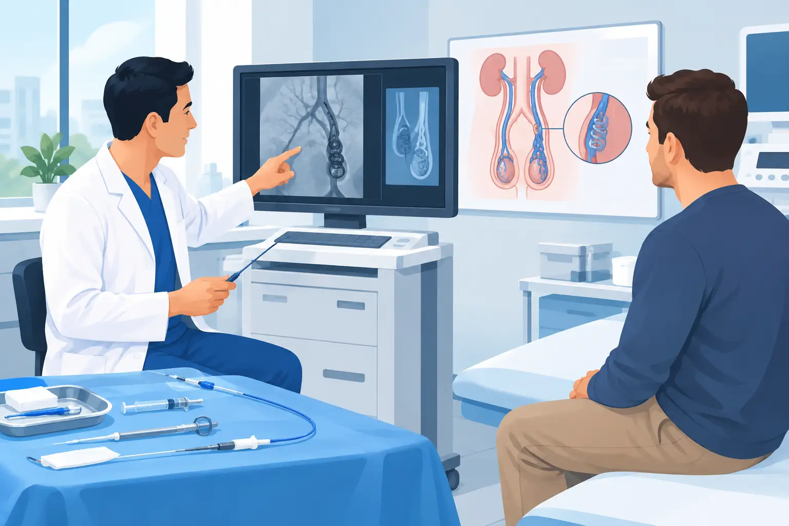
A varicocele can remain silent for years, or it can show itself as a feeling of heaviness in the scrotum, pain that gets worse when standing or playing sport, changes in the semen and concerns about fertility. Varicocele embolization without surgery is a treatment option for selected patients: it treats venous reflux from inside the vessel, through a small skin puncture and under radiological guidance.
It is not an automatic solution for every varicocele. The choice requires a careful clinical assessment, because the symptoms, the physical examination, the colour Doppler ultrasound, plans for having children and, when indicated, the semen analysis all matter. The aim is not simply to eliminate a dilated vein, but to stop the reflux that keeps the testicle congested with venous blood, while preserving the surrounding anatomy.
What is varicocele embolization without surgery?
A varicocele is an abnormal dilation of the veins that drain blood from the testicle. It is more common on the left, where the particular anatomy of the venous drainage can encourage increased pressure and backward flow of blood. In some men it has no consequences; in others it may be associated with pain, reduced testicular volume or abnormal semen parameters.
Embolization is an interventional radiology treatment. The doctor inserts a very thin catheter into a peripheral vein, usually through a puncture in the groin, arm or neck, depending on the anatomy and the clinical case. The catheter is guided with radiological imaging to the vein responsible for the reflux. Once the venous branches to be treated have been precisely identified, embolic materials such as metal coils and/or sclerosing agents are released to selectively close the diseased vessel.
Blood is thus redirected towards healthy venous pathways. No surgical incision is made in the scrotum or groin, the testicle is not handled directly and, in most cases, general anaesthesia is not needed. Saying “without surgery”, however, does not mean “without access”: there is still a small skin puncture, normally covered with a plaster at the end of the procedure.
When it may be indicated
A varicocele found by chance on ultrasound, with no symptoms and no clinically relevant abnormalities, does not always need treatment. Treating an imaging finding without a real indication is of no benefit to the patient.
Embolization may be considered when there is persistent pain or heaviness attributable to the varicocele, especially when it limits physical activity, work or quality of life. It may also be considered as part of the work-up for male infertility, if the varicocele is clinically significant and associated with an abnormal semen analysis. In adolescents and young adults, the decision requires particular attention to growth and testicular volume, with a dedicated andrological or urological assessment.
The relationship between varicocele and fertility is not the same for everyone. Any improvement in semen parameters after treatment takes time, because sperm production follows biological cycles lasting a few months. In addition, a couple's fertility also depends on the female partner's age and reproductive health. For this reason, when a couple is trying to conceive, the decision should be shared between the interventional radiologist, the andrologist or urologist and, when necessary, a fertility centre.
Tests before the procedure
The assessment starts with a consultation and a scrotal colour Doppler ultrasound, which is used to confirm the reflux, identify the side involved and study the venous anatomy. Semen analysis is relevant when the concern is fertility or abnormalities of the semen. Blood tests may be needed, including those for clotting and kidney function, depending on the medical history and the use of contrast medium.
It is essential to report anticoagulant or antiplatelet medication, known allergies to contrast medium, kidney disease, previous thrombosis and any current infections. Any change to your medication must never be decided on your own: it should be planned with the doctor who prescribes it and with the specialist who will perform the procedure.
How embolization is performed
The procedure is performed in an angiography suite, with monitoring of vital signs and under sterile conditions. After local anaesthetic at the access site, the patient remains awake and can talk to the team. In some cases light sedation may be helpful, but general anaesthesia is not usually required.
Contrast medium is injected through the catheter to show the veins and identify any collateral vessels. This step is central: precise imaging makes it possible to treat the vein involved and to reduce the risk of leaving active reflux pathways. The embolic material is chosen according to the anatomical configuration and the operator's experience. Based on experience and the clear evidence in the literature, I have chosen to use only cyanoacrylate, which guarantees the best results.
The duration varies, often between 30 and 60 minutes, but it can be longer if the venous anatomy is complex or collateral branches are present. After the catheter is removed, pressure is applied to the puncture site. Many patients go home the same day, after a short period of observation.
Recovery: what to really expect
In the first few hours there may be mild discomfort at the access site or a feeling of tension in the groin and scrotum. This is usually temporary and manageable with the pain-relief advice given by the doctor. A small local bruise is possible, especially if the puncture was made in the groin.
Generally, a quiet day is recommended, and strenuous physical exertion, the gym, demanding cycling and heavy lifting should be avoided for a few days. The exact timing depends on the access site, the type of work you do and how you progress individually. People with desk-based jobs can often return quickly; those with physically demanding work may need a few more days.
Pain relief is not always immediate. The treated venous system needs time to adapt, and mild discomfort in the following days does not necessarily mean the treatment has failed. Clinical and ultrasound follow-up makes it possible to confirm the reduction in reflux and relate the result to the patient's condition.
Benefits and limitations compared with surgery
The main advantage of embolization is percutaneous access: no traditional surgical incision, no stitches in the scrotum and, in most cases, no general anaesthesia. It is a targeted procedure that generally allows a quick recovery and going home the same day.
A further strength is the ability to study the venous network directly from the inside. This can be useful when collateral vessels are present or in some cases of persistence or recurrence after previous surgery. However, not all varicoceles can technically be embolized. Sometimes the vein cannot be reached safely, the anatomy is unfavourable or exposure to contrast medium makes another route preferable.
Surgery, in particular microsurgical varicocelectomy, remains an established and appropriate option in many situations. No technique is superior in absolute terms: both aim to stop venous reflux, but they differ in access, anaesthesia, recovery and technical profile. The right comparison is not between “modern” and “traditional”, but between the solutions best suited to the specific anatomy and the problem to be solved.
Risks and possible complications
Varicocele embolization is generally well tolerated when performed by an experienced team, but it is not free of risk. Bruising or bleeding at the puncture site, temporary pain, a reaction to contrast medium, inflammation of the treated vein or thrombosis may occur. More rarely, the embolic material may migrate, or a reflux pathway may persist and require further treatment.
The procedure also uses ionising radiation for image guidance. The dose is optimised according to the principle of the lowest necessary exposure, but this should be taken into account, especially in young patients or those who have already had many radiological examinations. Fever, significant or worsening pain, marked swelling or persistent bleeding after going home require prompt medical contact.
A targeted choice, not a shortcut
For anyone with a symptomatic varicocele or concerns about fertility, the useful question is not just “can I avoid an operation?”. It is understanding whether the varicocele is really the cause of the problem, whether treatment is indicated and which technique offers the best balance of effectiveness, invasiveness and safety in your own case. A specialist assessment with imaging and clear goals turns a complex decision into a well-reasoned pathway, with no automatic promises and no unnecessary treatment.
Want to know if it applies to you?
At the first consultation we review your clinical history and previous imaging together.
More articles
The information on this page is for general education and does not replace a medical consultation. The indication for any treatment must always be assessed individually.