
28 September 2026
Genicular artery embolization for knee osteoarthritis
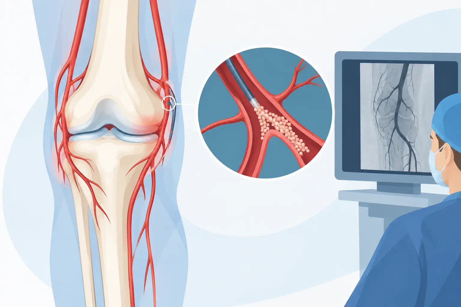
When osteoarthritis pain continues to limit stairs, walking, sleep and work despite physiotherapy and medication, the question is not just how to reduce it, but which treatment to use. Genicular artery embolization (GAE) for knee osteoarthritis is an interventional radiology procedure designed for some patients with persistent pain, with the aim of reducing hypervascularity and inflammation of the synovial membrane without a surgical incision.
It does not automatically replace a joint replacement, nor is it indicated for every osteoarthritic knee. It is an option to be assessed carefully, after correlating the symptoms, the clinical examination and the radiological images.
Why knee osteoarthritis can be so painful
Osteoarthritis is not just wear of the cartilage. In a painful knee there may be coexisting changes in the cartilage, the subchondral bone, the menisci and the synovium, the tissue that lines the inside of the joint. In some people the synovium remains inflamed and develops a network of small blood vessels that is more prominent than normal.
These vessels are not the sole cause of osteoarthritis, but they may help sustain inflammation and pain sensitisation. This is where embolization comes in: it does not work by rebuilding cartilage and does not correct major deformities, but selectively targets the arterial branches that supply the diseased areas of synovium.
This distinction is essential. A patient with mainly inflammatory pain, recurrent swelling and mild or moderate osteoarthritis may have a different profile from someone with severe malalignment, marked instability, joint locking or osteoarthritis that is already a candidate for joint replacement.
Genicular artery embolization: how it works
The genicular arteries are small arterial branches that surround the knee and contribute to the joint’s blood supply. During the procedure, the interventional radiologist introduces a very thin catheter into an artery, usually from the groin or the wrist. Using X-ray guidance with contrast medium, the catheter is advanced into the branches supplying the painful area.
Angiography makes it possible to recognise any areas of increased vascularity in the synovium. Once the target vessels have been identified, embolic microparticles are released selectively. The aim is to reduce the abnormal blood flow to the inflamed tissue while preserving the blood supply to healthy tissues.
Precision is not a technical detail: it is the necessary condition for achieving the desired result and limiting risks. This is why the angiographic study and knowledge of the vascular anatomy of the knee are an integral part of the treatment, not merely a preparatory step.
In most cases it is performed under local anaesthesia, sometimes with light sedation if needed. It does not require general anaesthesia or a surgical incision. At the end, a compression dressing is applied at the access site and, for suitable patients, they go home the same day.
What you feel during and after the procedure
The arterial access site is numbed with local anaesthetic. During the injection of contrast or particles, some patients may feel warmth or a temporary sensation of pressure in the knee area. After embolization there may be local soreness, mild swelling or a temporary increase in pain, generally manageable with the prescribed medication.
The benefit should not be judged the following day. The response is gradual and may take weeks, because the treatment acts on the vascular inflammatory process and does not produce an immediate mechanical effect.
Who may be a good candidate
Candidacy is determined during a specialist assessment, not just by reading an MRI or X-ray report. What matters is the exact location of the pain, how long it has lasted, the presence of effusion, the ability to walk, the treatments already tried and the person’s expectations.
In general, embolization may be considered in patients with symptomatic knee osteoarthritis and chronic pain that has not responded sufficiently to a well-conducted course of conservative treatment. This may include targeted physiotherapy, weight management when indicated, activity modification, medication or selected injections. It may be of interest to those who wish to avoid or postpone surgery, provided this choice is clinically reasonable.
It is not, however, an appropriate answer if the main problem requires mechanical or surgical correction. Severe instability, significant deformity, major structural limitation of the joint, active infection, certain vascular diseases and clotting disorders require particularly careful assessment or may be contraindications to the procedure.
The source of the pain also needs to be clarified. Knee pain may be due to osteoarthritis, but also to meniscal tears, tendinopathies, pain referred from the hip or spine, rheumatological conditions and other disorders. Treating the wrong vessel, or treating the knee when the source of the pain lies elsewhere, would not lead to the expected result.
Expected results: what the evidence says and what it does not promise
The available clinical studies on genicular embolization report, in selected patients, a reduction in pain and an improvement in function in the months after treatment. The data are encouraging particularly in mild-to-moderate osteoarthritis with an inflammatory component, but the literature continues to evolve: protocols, embolic materials, selection criteria and length of follow-up are not yet fully standardised.
For this reason it is wrong to present the procedure as a definitive cure for osteoarthritis. It does not regenerate cartilage, does not necessarily eliminate every symptom and does not guarantee that surgery will be avoided for ever. It can, however, offer a clinically significant reduction in pain and allow carefully selected people to regain daily activities, therapeutic exercise and independence.
The response varies. Some patients report a significant improvement, others a partial benefit and still others an insufficient effect. It is not possible to say in advance with certainty how long the result will last. Being transparent on this point is part of a correct indication.
Risks of embolization and follow-up after treatment
Like any endovascular procedure, embolization carries risks, although serious complications are uncommon when the indication and technique are appropriate. Haematoma or bleeding at the puncture site, reactions to the contrast medium, temporary pain, local skin changes due to non-target embolization and, rarely, more significant vascular problems may occur.
Before the procedure, allergies, kidney function, anticoagulant or antiplatelet therapy, diabetes, vascular history and the results of available tests should therefore be reviewed. Medicines that affect clotting must not be stopped on your own: their management is decided case by case with your doctor and the specialists involved.
After treatment, a short period of moderate activity is normally recommended, avoiding strenuous effort in the first few days. Recovery does not mean immobility: progressive movement and physiotherapy, if indicated, help turn the reduction in pain into a real functional improvement. Follow-up visits check how the symptoms are progressing, how the procedure was tolerated and whether the treatment pathway needs to be supplemented.
Embolization, injections or joint replacement: a tailored choice
Injections can be useful when the aim is to modulate joint pain and inflammation with an outpatient procedure, but results and duration depend on the drug used and the clinical picture. Physiotherapy remains central: improving quadriceps strength, movement control and load tolerance protects knee function, even when an interventional procedure is performed.
Knee replacement, on the other hand, is an established and often definitive treatment in cases of advanced osteoarthritis with severe impairment of quality of life. However, it requires surgery, a hospital stay and a more demanding recovery. Embolization is not an ideological competition with surgery: it is an intermediate option for the right patient, at the right time, with realistic goals.
The most useful decision starts from a concrete question: what is currently the main source of the pain, and which treatment can address it while preserving the anatomy and future options as much as possible? An accurate assessment makes it possible to answer without promising shortcuts, but without giving up targeted solutions when they are genuinely indicated.
Want to know if it applies to you?
At the first consultation we review your clinical history and previous imaging together.
More articles
The information on this page is for general education and does not replace a medical consultation. The indication for any treatment must always be assessed individually.