
22 October 2026
Greater trochanteric bursitis: how the ultrasound-guided injection works
Pain on the side of the hip that gets worse when lying on that side is often called trochanteric bursitis. Ultrasound shows that in many cases the gluteal tendons are involved, and lets the needle go exactly where it is needed. Here is how the ultrasound-guided injection works and what to expect.
Pain on the side of the hip
Pain on the outer side of the hip, right over the bone you can feel under the skin, that gets worse when lying on that side, climbing stairs or standing up after sitting for a long time. It is one of the most common reasons for a hip pain visit, especially in women aged 40 to 60. For years it was simply called trochanteric bursitis; today it is more often called greater trochanteric pain syndrome, because the pain can come from several structures close to one another. It should not be confused with hip osteoarthritis, which usually hurts in the groin, or with pain travelling down from the lower back: telling them apart is the first step towards the right treatment.
Bursa or tendons? Often both
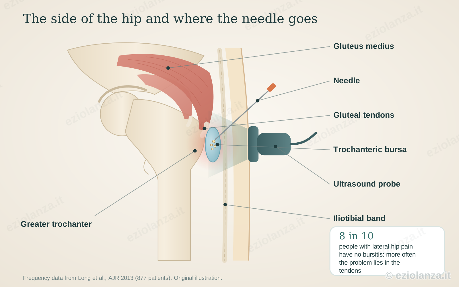
The tendons of the gluteus medius and minimus, the muscles that keep the pelvis level when we walk, attach to the greater trochanter, the bony prominence on the outside of the femur. Between these tendons and the iliotibial band, a strong sheet of tissue running over them, lie small gliding sacs called bursae. When the tendons are overloaded and inflamed, the nearby bursa can fill with fluid. In a study of 877 people with lateral hip pain assessed with ultrasound, only 20% actually had bursitis; about half had a problem in the gluteal tendons and nearly three in ten a thickened iliotibial band. Knowing this changes treatment: ultrasound shows what is really there, and therefore where the needle should go.
How the ultrasound-guided injection is done
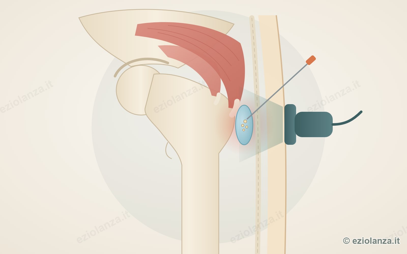
You lie on your healthy side. After cleaning the skin, I place the ultrasound probe on the side of the hip and the screen shows the trochanter, the tendons and the bursa. A thin needle goes in a few centimetres from the probe and I follow it in real time as it advances, up to the bursa or the space around the affected tendon. There I deposit a local anaesthetic and a corticosteroid, and I see the drug spread where it is needed. The corticosteroid must not be injected into the tendon, which would be weakened: ultrasound makes it possible to avoid this. It takes about fifteen minutes, with no radiation, and you walk home. If the pain disappears straight away thanks to the anaesthetic, that also confirms it was coming from there.
What the studies show
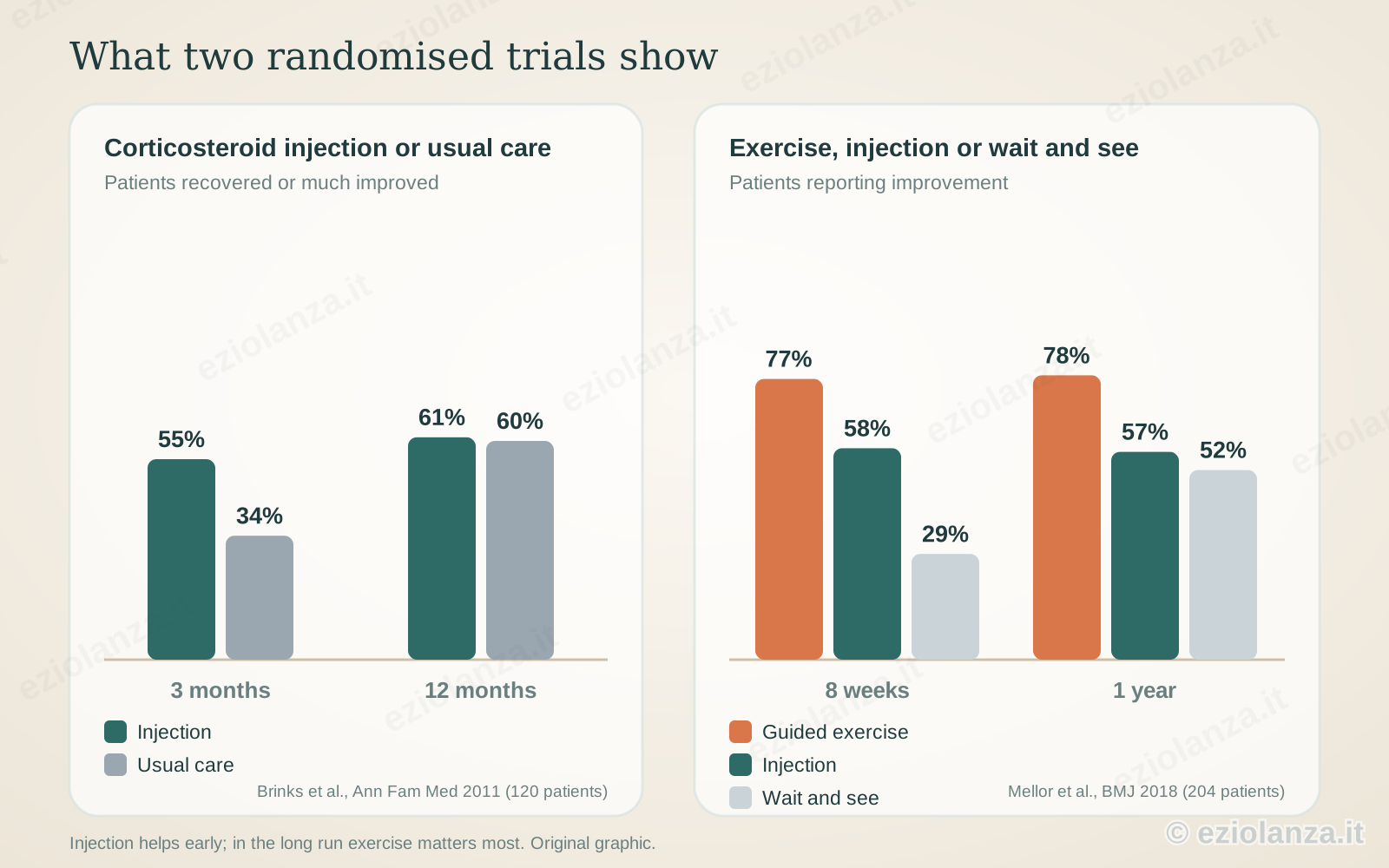
In a Dutch randomised trial of 120 patients treated by their family doctor, at three months 55% of those who received a corticosteroid injection had recovered or were much improved, compared with 34% of those on usual care. At one year, however, the two groups were the same, around 60%. The Australian LEAP trial, published in the BMJ in 204 people with gluteal tendinopathy, compared an exercise and education programme, a single injection and waiting: at eight weeks 77% of the exercise group reported improvement, 58% of the injection group and 29% of those who waited; at one year exercise was still ahead. The message is clear: an injection brings quick relief, but a lasting result is built with exercise.
Why with ultrasound
In a BMJ study, bursa injections guided with X-rays did not give better results than freehand ones. The advantage of ultrasound lies elsewhere. First in diagnosis: it shows whether the problem is the bursa, the tendon, a partial tear or a calcification, and helps recognise people who instead have a problem in the joint or the spine. Then in precision: the deeper bursae, under the gluteus medius, and hips with more subcutaneous tissue are hard to reach blind. Finally in safety: the needle is visible, injecting the corticosteroid into the tendon is avoided and no radiation is used.
After the injection: the role of exercise
The corticosteroid calms the inflammation, but it does not strengthen the tendons. That is why, after a few days, I always recommend a gluteal exercise programme, ideally guided by a physiotherapist, and a few everyday habits: do not cross your legs, do not stand for long leaning on one hip, sleep on your side with a pillow between your knees. The most recent reviews of the evidence, from 2024 and 2025, point to exercise as the core treatment, with the injection as a help to get through the most painful phase and be able to do the exercises.
If the pain comes back
Corticosteroid injections can be repeated, but with judgement and no more than two or three times a year in the same place. When the pain comes back despite exercise and injections, it is worth rechecking the tendons with ultrasound. Shockwave therapy has also been studied, with a short-term benefit in studies of still limited quality, as have other types of injection, for which data are still scarce. In cases that respond to nothing, embolization of the small inflamed vessels around the trochanter can be considered, which I discussed in another article, while surgery is reserved for a few selected cases, such as complete tendon tears.
In short
Pain on the side of the hip is common and is almost always treated without surgery. Ultrasound helps to understand whether the problem lies in the bursa, the tendons or elsewhere, and guides the needle exactly to the right spot. The injection brings relief within a few weeks; to make it last, exercise is needed. At the visit we look at your symptoms and the ultrasound together, and choose the path that suits you best.
Treatments discussed in this article
Scientific sources
- Long SS, Surrey DE, Nazarian LN. Sonography of greater trochanteric pain syndrome and the rarity of primary bursitis. AJR Am J Roentgenol. 2013;201(5):1083-1086.
- Brinks A, van Rijn RM, Willemsen SP, et al. Corticosteroid injections for greater trochanteric pain syndrome: a randomized controlled trial in primary care. Ann Fam Med. 2011;9(3):226-234.
- Mellor R, Bennell K, Grimaldi A, et al. Education plus exercise versus corticosteroid injection use versus a wait and see approach on global outcome and pain from gluteal tendinopathy: prospective, single blinded, randomised clinical trial. BMJ. 2018;361:k1662.
- Cohen SP, Strassels SA, Foster L, et al. Comparison of fluoroscopically guided and blind corticosteroid injections for greater trochanteric pain syndrome: multicentre randomised controlled trial. BMJ. 2009;338:b1088.
- Kjeldsen T, Hvidt KJ, Bohn MB, et al. Exercise compared to a control condition or other conservative treatment options in patients with greater trochanteric pain syndrome: a systematic review and meta-analysis of randomized controlled trials. Physiotherapy. 2024;123:69-80.
- Wang SQ, Guo NY, Liu W, et al. Effect of conservative treatment on greater trochanteric pain syndrome: a systematic review and network meta-analysis of randomized controlled trials. J Orthop Surg Res. 2025;20(1):126.
- Rhim HC, Shin J, Beling A, et al. Extracorporeal shockwave therapy for greater trochanteric pain syndrome: a systematic review with meta-analysis of randomized clinical trials. JBJS Rev. 2024;12(8).
Want to know if it applies to you?
At the first consultation we review your clinical history and previous imaging together.
More articles
The information on this page is for general education and does not replace a medical consultation. The indication for any treatment must always be assessed individually.