
1 October 2026
Ultrasound-guided treatment of tennis elbow
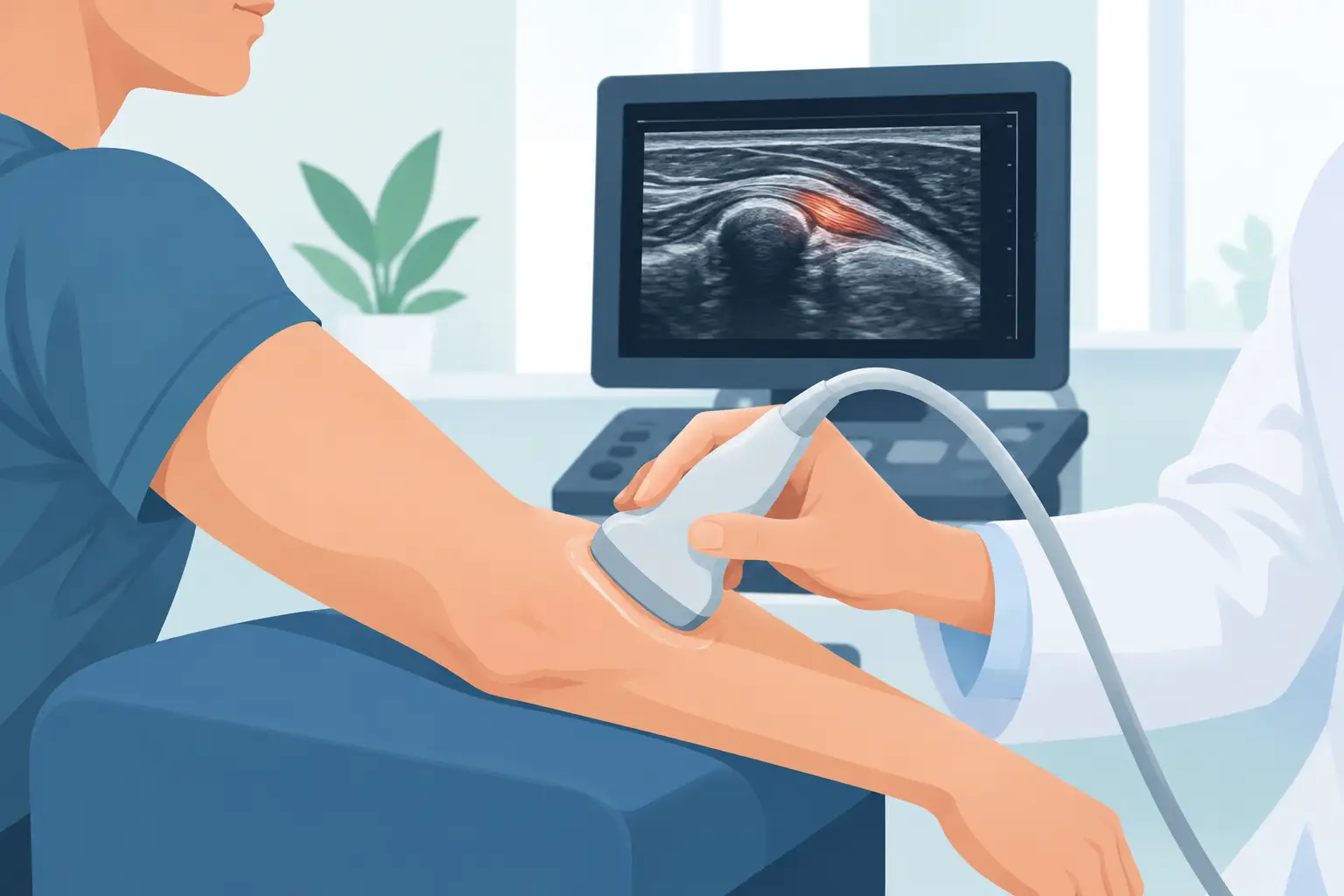
A pinpoint pain on the outer side of the elbow can make it hard to shake hands, lift a bag or use a mouse for hours at a time. Ultrasound-guided treatment of lateral epicondylitis (tennis elbow) is not a shortcut that suits everyone, but it can be a targeted option when pain persists despite relative rest, well-conducted physiotherapy and correction of the movements that overload the tendon.
Ultrasound makes it possible to see the area to be treated in real time: not just the spot where the patient feels pain, but the common extensor tendon, its attachment to the epicondyle and the surrounding tissues. This changes the way an injection or percutaneous procedure is planned: the aim is not to “give an injection in the elbow”, but to reach a specific anatomical abnormality precisely, avoiding nerves, vessels and healthy structures.
What epicondylitis is and why it can become chronic
Lateral epicondylitis (tennis elbow) mainly affects the tendons that allow you to extend the wrist and fingers. It does not only affect people who play tennis: it is common in people who do manual work, use vibrating tools, spend many hours at the computer, do gardening or repeatedly lift weights.
In its early stages, the problem may be linked to temporary overload. When the pain lasts for months, however, the tendon tissue can show signs of tendinopathy: disorganised fibres, thickening, small tears and increased vascularity. At this stage it is not necessarily a simple “inflammation”. This is why repeated and unselective use of anti-inflammatory drugs or cortisone injections does not always solve the problem at its root.
A specialist consultation also helps avoid automatically attributing every pain on the outer side of the elbow to epicondylitis. Neck problems, nerve compression, joint problems or pain referred from the shoulder can produce similar symptoms. An accurate diagnosis comes before any treatment.
When to consider ultrasound-guided treatment of tennis elbow
Ultrasound-guided treatment is considered mainly when pain limits work, sport or daily activities and does not improve after an adequate course of conservative treatment. There is no threshold that is the same for everyone: what matters is how long the symptoms have lasted, where the lesion is, the degree of functional limitation, the treatments already tried and the person’s realistic expectations.
In many cases, physiotherapy and progressive strengthening exercises for the forearm muscles remain the foundation of treatment. Modifying the load does not mean completely immobilising the arm, but dosing painful activities and reintroducing them gradually. A brace can help at certain times, but it does not replace restoring the tendon’s ability to tolerate effort.
An ultrasound-guided procedure can complement this pathway when ultrasound shows a localised tendinopathy and rehabilitation alone has not provided sufficient benefit. One point needs to be clear: the treatment does not remove the need for rehabilitation. It can reduce pain or stimulate a healing process, but the result also depends on how the tendon is managed in the following weeks.
What ultrasound shows before the procedure
Musculoskeletal ultrasound is a dynamic examination. It allows the painful elbow to be compared with the other side, the thickness and echotexture of the tendon to be assessed, and calcifications, degenerative areas and abnormal vessels to be identified. During the scan it is also possible to check whether the pain is reproduced by pressing on the abnormal spot or by specific movements.
This information guides the choice. If the tendon looks largely preserved and the problem is recent, continuing with a conservative programme may be more appropriate. If, on the other hand, there are persistent focal changes, ultrasound guidance makes it possible to plan a truly selective percutaneous procedure.
Ultrasound should not be seen as an isolated image to be interpreted without the patient. A tendon finding may also be present in people with no pain. The clinical decision comes from combining symptoms, examination, arm function and imaging.
Which procedures can be performed under ultrasound guidance
There is no single ultrasound-guided injection for epicondylitis. The solution must be chosen case by case, explaining transparently what benefit can reasonably be expected and what the limits of the available evidence are.
In some situations a peritendinous injection may be indicated, that is, around the tendon rather than into it, with selected drugs to control a particularly intense painful phase. Cortisone can provide short-term relief, but requires caution in chronic tendinopathies: repeated or incorrectly placed injections can weaken the tissue and do not guarantee a lasting benefit.
Where there is localised tendon degeneration, the specialist may consider percutaneous needling or fenestration techniques. Using a fine needle introduced under ultrasound control, small passes are made through the diseased area with the aim of stimulating a repair response. Depending on the clinical picture and the indication, autologous blood or platelet concentrates may be added. These options are not equally effective in every patient and should not be presented as guaranteed definitive cures.
The choice also depends on whether calcifications are present, how deep the lesion is, the medicines being taken and conditions such as diabetes, clotting disorders or anticoagulant therapy. These aspects must be discussed before the procedure, without stopping medicines prescribed by other specialists on your own initiative.
How the ultrasound-guided procedure is carried out
The treatment is generally performed as an outpatient procedure. The patient lies down or sits with the arm resting in a stable position. After the skin has been disinfected, the ultrasound scanner is used to locate the target and plan the needle’s path.
The needle is seen in real time as it reaches the planned site. This is the essential difference compared with an injection performed only on the basis of external anatomical landmarks: the doctor can check where the tip is and monitor how any drug is distributed. When necessary, local anaesthesia is used; general anaesthesia is not required and, in most cases, the patient goes home the same day.
The actual interventional part is short, but the consultation and preparation take the time needed to review tests, indications and informed consent. A well-performed treatment is not just a technical procedure: it is also a shared decision about goals, alternatives and recovery.
Pain after treatment, recovery and expected results
After a percutaneous procedure you may feel local soreness for a few days. With needling techniques, a temporary flare-up can be part of the tissue’s response and does not automatically mean failure. Ice, if recommended, and a temporary reduction in loads can be helpful.
Return to light activities is often quick, but physically demanding work, racket sports or weight training require a gradual return. Timings vary according to the procedure and the condition of the tendon. After a simple injection, the advice may be different from that after tendon fenestration. Forcing grip or wrist extension too early can keep the overload cycle going.
Improvement is not always immediate. If the aim is to promote recovery of a degenerated tendon, the result is assessed over weeks or months, together with function: grip strength, ability to work, sleep quality and return to desired activities. A clinical and ultrasound follow-up, when indicated, helps check progress without treating the image as the only measure of success.
Risks, limits and alternatives to be aware of
Ultrasound guidance increases precision and safety, but does not eliminate risk. Temporary pain, bruising, very rare infection, reaction to the drug or lack of benefit may occur. The specific risk depends on the technique used and on individual circumstances.
Even a technically correct treatment may not bring the hoped-for improvement if the initial diagnosis was incomplete, if the tendon continues to be overloaded or if the pain has several components. In a proportion of cases with resistant symptoms and significant functional impairment, it may also be appropriate to discuss surgical options. Surgery is not the automatic next step after a single injection fails, nor should an ultrasound-guided procedure delay it when there is a clear indication.
For anyone who has lived with pain on the outer side of the elbow for some time, the useful starting point is an assessment that looks at the tendon, the movement that overloads it and the practical goal to be regained. Treating with precision means choosing the minimum intervention needed, at the right time, and combining it with a concrete programme to start using the arm with confidence again.
Want to know if it applies to you?
At the first consultation we review your clinical history and previous imaging together.
More articles
The information on this page is for general education and does not replace a medical consultation. The indication for any treatment must always be assessed individually.