
27 September 2026
Ultrasound-guided lavage for shoulder calcification
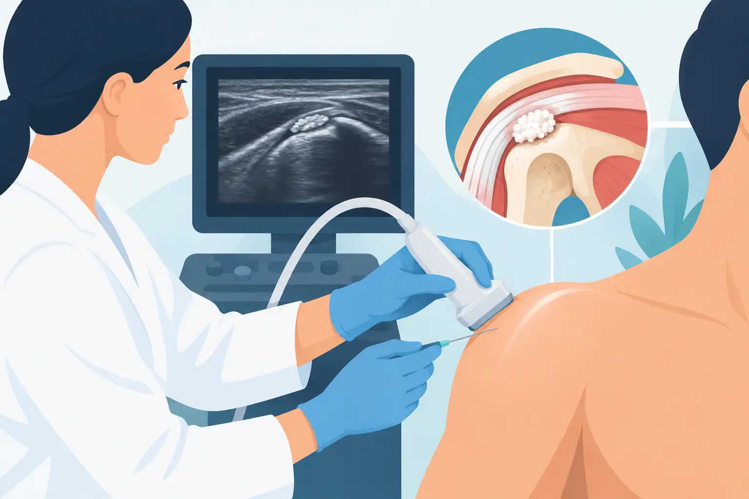
A sharply localised but very intense pain in the upper arm, especially at night or when trying to raise the arm, may be caused by calcification of the rotator cuff tendons. Ultrasound-guided lavage (barbotage) for shoulder calcification is a minimally invasive treatment that breaks up and aspirates the calcium deposit through small needles, with every step monitored by ultrasound. It is not an automatic solution for every painful shoulder, but it can be very effective when the diagnosis and indication are correct.
What is shoulder calcification?
Calcific tendinitis of the shoulder, also called calcific tendinopathy, is the build-up of calcium crystals inside a tendon, most often the supraspinatus tendon. This tendon is part of the rotator cuff, the group of muscles and tendons that stabilises the shoulder and allows the arm to move.
The deposit is not simply the result of too much calcium in the diet or in the blood. The causes cannot always be identified, and local factors in the tendon, overuse, age and changes in its blood supply may all contribute. The calcification may remain silent for years, be found by chance on an X-ray, or cause sudden, disabling pain.
In the most inflammatory phase, the pain can make everyday actions difficult: getting dressed, combing your hair, driving, sleeping on the affected side or reaching for something on a shelf. In some cases inflammation of the subacromial bursa accompanies the deposit and amplifies the symptoms.
When ultrasound-guided lavage is indicated
Before ultrasound-guided lavage of a shoulder calcification is proposed, it is essential to confirm that the deposit is really responsible for the pain. Ultrasound makes it possible to assess the location, size and consistency of the calcification, the rotator cuff tendons, any bursitis and the presence of associated lesions. In selected situations, an X-ray or MRI completes the picture.
The treatment is considered mainly when pain persists despite relative rest, targeted physiotherapy and medication prescribed by a doctor, or when the functional limitation is significant. It is particularly suitable for calcifications that can be reached with a needle and whose characteristics allow them to be broken up and aspirated.
Not all calcifications are the same. A very small, hard deposit that causes no symptoms does not necessarily require a procedure. Conversely, a very stiff shoulder may need a specific treatment pathway for adhesive capsulitis, even if a calcification is also present. If the pain comes mainly from a tendon tear, arthritis or another cause, lavage does not solve the underlying problem.
The clinical decision must therefore start from the symptoms and examination of the shoulder, not from the imaging alone. Treating a calcification that is visible but not painful offers no proportionate benefit.
How ultrasound-guided lavage is performed
The procedure is carried out as an outpatient, generally without general anaesthesia and without a surgical incision. The patient is positioned so that the shoulder is exposed and the area is carefully disinfected. After local anaesthesia of the skin and superficial tissues, the doctor inserts one or two fine needles into the deposit under continuous ultrasound guidance.
Ultrasound is not only used to find the calcification. It allows the needle tip to be seen in real time, important structures to be avoided and precise work to be carried out inside the deposit. Saline is injected to loosen and break up the calcific material, which is then aspirated whenever possible. This technique is also known as barbotage or percutaneous lithoclasy.
The material removed may look whitish and paste-like, but the calcification cannot always be removed completely. The aim is not to achieve a perfectly clean X-ray at all costs: it is to reduce the irritating burden of the deposit and help movement return with less pain.
At the end, if indicated, an ultrasound-guided injection into the subacromial bursa may be performed to control reactive inflammation. This choice depends on the ultrasound findings, the symptoms and the person's clinical condition. After a short period of observation, the patient goes home the same day.
How long it takes and what you feel during treatment
The duration varies with the position and consistency of the calcification, but the procedure generally takes about 20-30 minutes. Local anaesthesia limits the discomfort of the needles going in; during the lavage you may feel pressure or a sense of tension in the shoulder. Ongoing communication during the treatment helps adapt each step to what the patient can tolerate.
Recovery after calcification lavage
In the first 24-72 hours, local pain or a temporary increase in soreness is common. This may be due to the handling of the deposit and the inflammatory response of the surrounding tissues. The doctor gives individual advice on the use of ice, painkillers or anti-inflammatories, taking into account allergies, current medication and general health.
It is not usually necessary to immobilise the shoulder completely. Relative rest in the first few days is sensible, avoiding strain, lifting, sport and repetitive overhead movements. The return to normal activities depends on the kind of work you do and on your response to treatment: for desk-based work it can be quick, whereas physical work or sport calls for more caution.
Physiotherapy often plays a useful role after the acute phase. Regaining mobility, gradually strengthening the rotator cuff and correcting any compensatory movements of the shoulder blade all help to make the result more lasting. Lavage treats the deposit, but it does not always replace rehabilitation when there is stiffness, weakness or altered movement patterns at work or in sport.
Expected results and real limitations
Many patients report a significant reduction in pain and improved function in the following weeks. The timescale, however, is not the same for everyone. A soft calcification that is already being reabsorbed tends to respond differently from a compact, long-standing deposit; in addition, bursitis, joint stiffness or coexisting tendon lesions can slow recovery.
Improvement may be gradual rather than immediate. Even when residual deposit is still visible on imaging, the patient may feel well and move the shoulder without restriction. Conversely, complete disappearance on imaging is not the only useful measure: what matters most is getting back to daily activities and to good-quality sleep.
Sometimes lavage does not manage to aspirate a significant amount of calcium, or the benefit is only partial. In these cases the diagnosis, the characteristics of the deposit and the alternative treatments are reassessed. A second procedure may be appropriate in selected circumstances, but it should not become automatic.
Risks and precautions
Ultrasound-guided lavage is a percutaneous procedure that is generally well tolerated, but it is not free of risk. Post-procedure pain, bruising, local bleeding and temporary inflammation are the most common events. Infection, reactions to medication and injury to nearby structures are rare, but must be explained before consent is given.
Ultrasound guidance reduces uncertainty because it shows the target and the needles throughout the procedure, without using ionising radiation. It does not, however, eliminate every risk. Anticoagulant or antiplatelet therapy, poorly controlled diabetes, allergies, skin infections at the access site and specific medical conditions all require careful assessment beforehand.
Lavage, shockwave therapy or surgery?
Focused shockwave therapy can be a non-invasive option for some calcific tendinopathies, especially when the pain is manageable and there is no indication to remove the deposit straight away. It does, however, require several sessions and the response may be gradual. There is no single best option: it depends on the stage of the condition, the shape of the calcification and the patient's goals.
Arthroscopic surgery is considered in a minority of cases, for example when symptoms persist despite well-conducted treatment or when there are associated problems that require an operation. Compared with surgery, ultrasound-guided lavage preserves the anatomy, involves no surgical incisions or general anaesthesia and allows you to go home the same day. Its limitation is that it cannot treat every shoulder condition and does not guarantee complete aspiration of every deposit.
For anyone who has been living with shoulder pain for weeks or months, the most useful question is not which treatment is fastest in the abstract, but which procedure is proportionate to their own problem. A specialist assessment with ultrasound can turn a report of calcification into a concrete, targeted and realistic path towards recovering movement.
Want to know if it applies to you?
At the first consultation we review your clinical history and previous imaging together.
More articles
The information on this page is for general education and does not replace a medical consultation. The indication for any treatment must always be assessed individually.