
30 September 2026
How does an ultrasound-guided injection work?
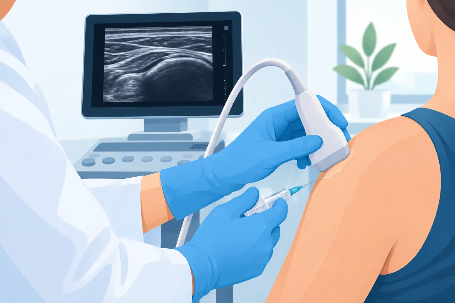
When pain in the shoulder, knee, hip, wrist or foot persists despite rest, medication or physiotherapy, the question is not only which drug to use, but where to deliver it. Understanding how an ultrasound-guided injection works helps with exactly this: assessing whether a local, targeted treatment without a surgical incision might be suitable in your case.
Ultrasound allows the specialist to see tendons, bursae, muscles, joints, effusions and superficial nerve structures in real time. The needle is therefore not inserted on the basis of anatomical landmarks or palpation alone: it is guided to the site that needs to receive the treatment. This difference can be relevant both to the effectiveness and to the safety of the procedure.
How an ultrasound-guided injection works, step by step
An ultrasound-guided injection is an outpatient procedure in which a drug or therapeutic substance is delivered through a fine needle under ultrasound control. It does not require surgical incisions or general anaesthesia. In most cases the patient goes home the same day.
The first step is always the clinical assessment. Pain alone does not identify a diagnosis: pain on the outer side of the hip may come from the gluteal tendons or the trochanteric bursa; wrist pain may involve the joint, a tendon sheath or a nerve. The specialist takes the history of the symptoms, examines the affected area and uses ultrasound to check that the structure seen is consistent with the problem reported.
If the injection is appropriate, the patient is positioned so that the area to be treated is accessible and stable. The skin is carefully disinfected, sterile ultrasound gel is applied or the probe is protected with a sterile cover, and the needle is inserted. On the screen, the specialist follows the path of the needle and its tip all the way to the target.
The drug is injected slowly. Depending on the indication, a local anaesthetic, a corticosteroid, hyaluronic acid or other substances included in the treatment plan may be used. There is no one-size-fits-all injection: the choice depends on the diagnosis, the site, the inflammation present, the medical history and any treatments already received.
After the procedure, which is generally short, a small dressing is applied. The patient receives personalised advice on which activities are allowed, on the possible use of ice and on the warning signs for which to contact the doctor.
Why ultrasound makes the difference
Ultrasound is not just an extra check. It makes it possible to confirm the position of the needle and direct the drug to the intended spot: inside a joint, into a bursa, around a tendon or close to a nerve structure, when the indication calls for it.
This approach is particularly useful in anatomical sites that are small, deep or crowded with delicate structures. In thumb osteoarthritis, for example, the trapeziometacarpal joint is small; in the shoulder, bursitis needs to be distinguished from a tendon problem; in the hip and trochanteric region, it is useful to avoid treating a painful area generically without identifying the tissue involved.
Ultrasound guidance also makes it possible to avoid, where possible, blood vessels, nerves and other structures that should not be crossed. It does not eliminate every risk, but it makes the procedure more controlled and reproducible than an injection performed without direct visualisation.
Which problems it may be considered for
Ultrasound-guided injections are used in many musculoskeletal conditions, but they are not automatically the first choice. They may be considered, for example, for shoulder or hip bursitis, epicondylitis, joint pain from osteoarthritis of the knee, hip, wrist or thumb, plantar fasciitis, tenosynovitis and some selected forms of low back or peripheral pain.
The indication changes depending on the mechanism of the pain. Significant acute inflammation may benefit from a local anti-inflammatory drug, whereas in osteoarthritis the aim may be to temporarily improve pain and function so that movement and therapeutic exercise become possible. In the case of shoulder calcification, a simple injection may not resolve the calcium deposit: when appropriate, ultrasound-guided percutaneous lavage of the calcification, also called lithoclasty or barbotage, may be considered.
Physiotherapy also often keeps a central role. Reducing pain does not automatically correct the muscle weakness, overload, stiffness or repetitive movements that contributed to the problem. An injection can create a useful window to regain movement and carry out more effective rehabilitation, but the result depends on the diagnosis and on the overall programme.
What you feel during and after the procedure
The sensation varies. The skin puncture may cause brief discomfort, similar to a blood test, while the injection may cause pressure, burning or a feeling of local tightness for a few seconds. In sensitive areas or where there is marked inflammation the discomfort may be greater, but the procedure is normally well tolerated.
Over the following hours, temporary local pain may occur. After a corticosteroid, some people experience a temporary flare-up for one or two days before any anti-inflammatory effect. The local anaesthetic, if used, can provide immediate but short-lived relief: this should not be taken as the final result of the treatment.
Post-procedure advice depends on the area treated and the drug used. It is often recommended to avoid intense physical activity and repetitive loading for 24–48 hours, without imposing unnecessary immobility. Driving, working at a computer or walking may be possible in many cases, but a procedure on the foot, the knee or the dominant limb requires individual practical considerations.
Real benefits and limits to be aware of
The main advantage is precision: treating a structure identified on ultrasound, with real-time visualisation, without a surgical incision and usually without general anaesthesia. For many patients this means an outpatient pathway, shorter timescales and a return to normal activities compatible with the site treated.
However, precision does not mean a guaranteed cure. An injection may give significant, partial or no relief. How long the effect lasts varies greatly: it depends on the condition, the stage of the disease, the substance injected, subsequent strain and the presence of factors such as excess weight, diabetes, repetitive work or biomechanical abnormalities.
Repeating injections without reassessing the diagnosis is not a good strategy. In particular, repeated use of corticosteroids in some sites may have limitations related to tissue quality and to local or systemic effects. Decisions need to be made case by case, avoiding both the idea that an injection is always harmless and the fear that it is necessarily a treatment to be avoided.
Risks and contraindications
Like any needle procedure, an ultrasound-guided injection carries risks, although these are generally uncommon when it is performed in appropriate conditions. Temporary pain, a small haematoma, a vasovagal reaction, infection, bleeding or reactions to the drug may occur. More serious complications are rare, but they must be explained before consent is given.
Anticoagulant or antiplatelet therapy does not automatically make the procedure impossible, but it requires careful planning. You should not stop medicines on your own initiative: the decision depends on the specific drug, your personal thrombotic risk and the type of injection planned. Diabetes, allergies, skin infections at the access site, fever, pregnancy and previous surgery also deserve assessment beforehand.
After the treatment, you should promptly report fever, increasingly severe pain, spreading redness, discharge from the puncture site, significant swelling or unusual symptoms. These events are uncommon, but recognising them without delay is part of a safe pathway.
How to prepare for an injection consultation
It is useful to bring previous tests, such as ultrasound scans, X-rays, MRI scans, reports and an up-to-date list of your medicines. These documents guide the assessment, but they do not replace the clinical and ultrasound examination carried out at the time of the consultation.
It is a good idea to wear clothing that makes it easy to expose the affected area, and to describe precisely where it hurts, for how long, which movements trigger the symptom and which treatments have already helped or failed. Apparently simple information, such as night-time shoulder pain or being unable to open a jar because of thumb pain, helps define the treatment goal.
The right question is not whether to have an injection at all costs, but whether that treatment targets the most likely cause of your pain. A well-indicated procedure, performed under ultrasound guidance and included in a realistic treatment plan, can reduce symptoms and restore function without turning a complex problem into a simple promise.
Want to know if it applies to you?
At the first consultation we review your clinical history and previous imaging together.
More articles
The information on this page is for general education and does not replace a medical consultation. The indication for any treatment must always be assessed individually.